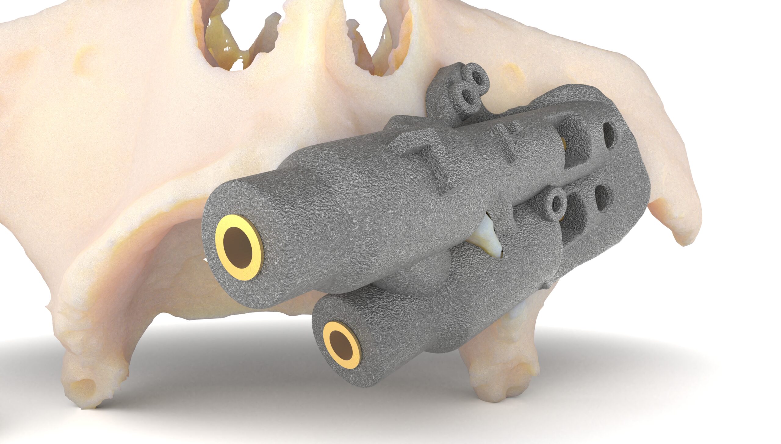
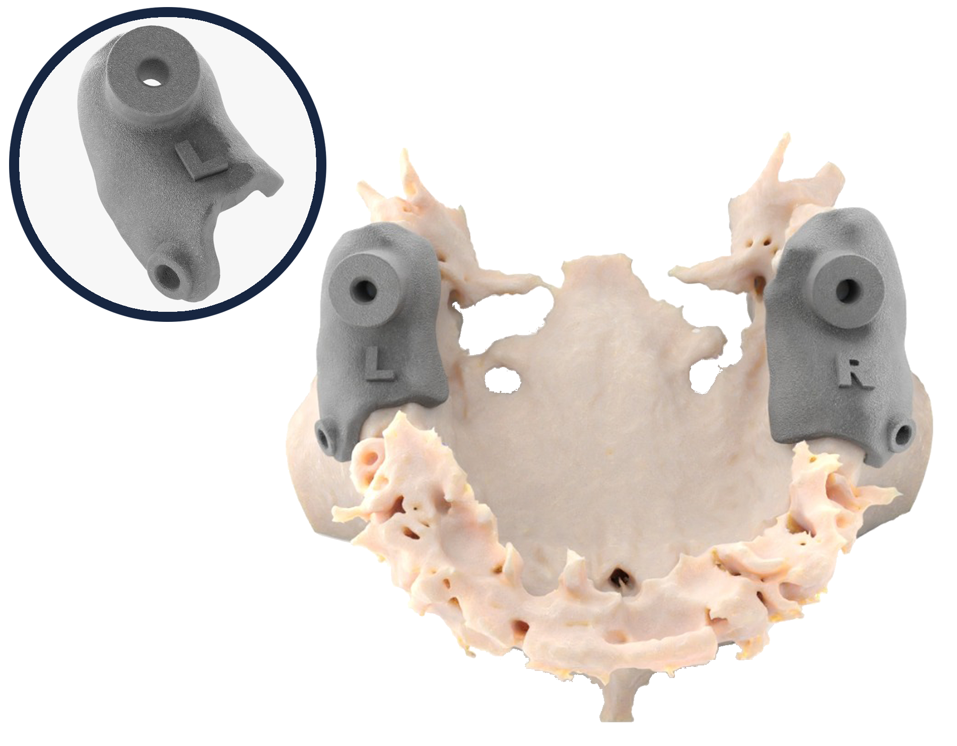
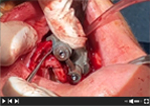
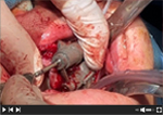
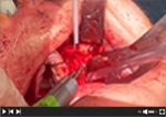
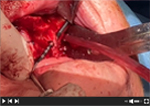
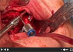
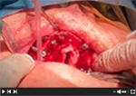
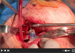
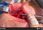
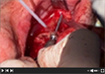
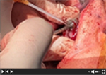
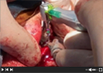
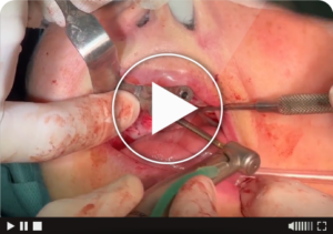
A CT scan allows planning and fabrication of surgical guides and the patient’s biomodel for precise, safe and personalized surgery for zygomatic and pterygoid treatments.
Advantages:
Treatment planning based on bone and/or prosthesis.
Anatomical and precise adjustment of the guides to the jaw.
High precision of the drilling.
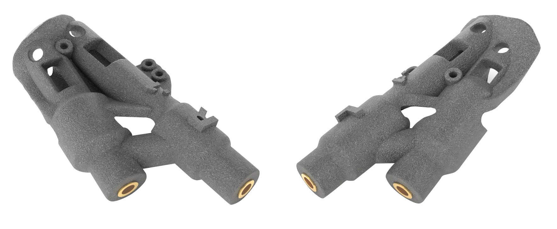
Metal guided tubes 9mm. for precise drilling adjustment.
Reduces operating room time.
Fixation of the guides with osteosynthesis screws.
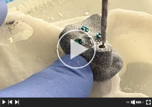
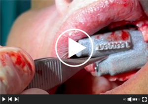
Demonstrative video surgical protocol zygoma surgical guides
Demonstration video: Demonstrative video surgical protocol Pterygoid Full Guided surgical guides
Demonstration video surgical protocol for Pterygoid Pilot surgical guides
Video procedure for planning and manufacturing of zygomatic guides.
Video planning procedure pterygoid guides.
Zygomatic & pterygoid guide brochure
CT procedure surgical guides zygomas and pterygoids
Case report Dr. Alejandro Gutiérrez Jiménez
Main benefits of the zygoma guides
• Milling precision at the three key points of the maxilla: the crest and the malar entry and exit.
• Facilitates the placement of two implants in small malars.
• Planning according to the diagnostic wax-up (double CT technique).
– Easy provisionalization on the day of the surgery. – Reduction of prosthodontist’s time.
Main benefits of the pterygoid guides
• Ensure correct drilling into the pterygoid bone.
– Correct angulation of the implant. – Anticipate during the planning of possible angled abutments. – Plan implant bi-corticalization for optimal primary stability.
Surgery with Zygoma (Quad) implants and surgical guides.
An expert maxillofacial surgeon teaches another colleague all the secrets of surgery; tips and keys to safe and accurate surgery.
12 videos showing each step of the surgery.
Video 1.
Incision and opening of the flap.
Video 2.
Adjustment and fixation with osteosynthesis screws of the surgical guide in the maxilla Q2.
Video 3.
Drilling and subsequent removal of the Q2 guide.
Video 4.
Preparation of the alveolar canal and bone crest for proper placement of the Q2 zygomatic implant.
Video 5.
Measurement of the alveolus, selection of the size, and insertion of the Q2 implant.
Video 6.
Prosthetic implant axis, final verification, and disconnection of the Q2 abutment.
Video 12.
Suture, placement of the Multi-unit abutments and healing caps.
Video 11.
Prosthetic axis of the implant, final verification, and disconnection of the Q1 abutment.
Video 10.
Measurement of the alveolus, selection of the size, and insertion of the Q1 implant.
Video 9.
Preparation of the alveolar canal and bone crest for the correct placement of the Q1 zygomatic implant.
Video 8.
Drilling and subsequent removal of the Q1 guide.
Video 7.
Adjustment and fixation with osteosynthesis screws of the surgical guide in the maxilla Q1.
Quad zygoma surgery with Kune surgical guides
Dr. Jorge Giner Díaz MD, DDS, FEBOMS
II Quad Zygoma Surgery with Kune Surgical Guides
Dr. Jorge Giner Díaz MD, DDS, FEBOMS
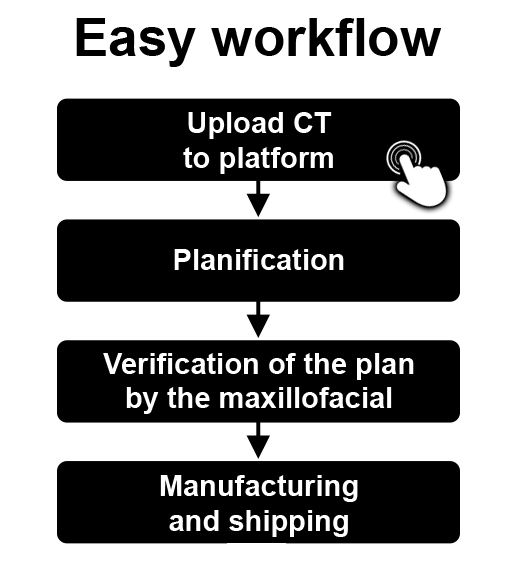
Workflow
UPLOAD CT TO KUNE´S IMPLANT CLOUD
The maxillofacial uploads the patient's CT scan to the cloud
TREATMENT PLANNING
Kune implants verify the quality of the CT scan
A zygomatic implant planning is performed based on the maxillary bone and the diagnostic wax-up (in case the maxillofacial has uploaded a double CT scan)
Kune sends a plan to the maxillofacial for verification and approval. The practitioner can make the necessary changes in remote control
MANUFACTURING AND SHIPPING
Manufacturing of surgical guides and biomodel
Customer shipment
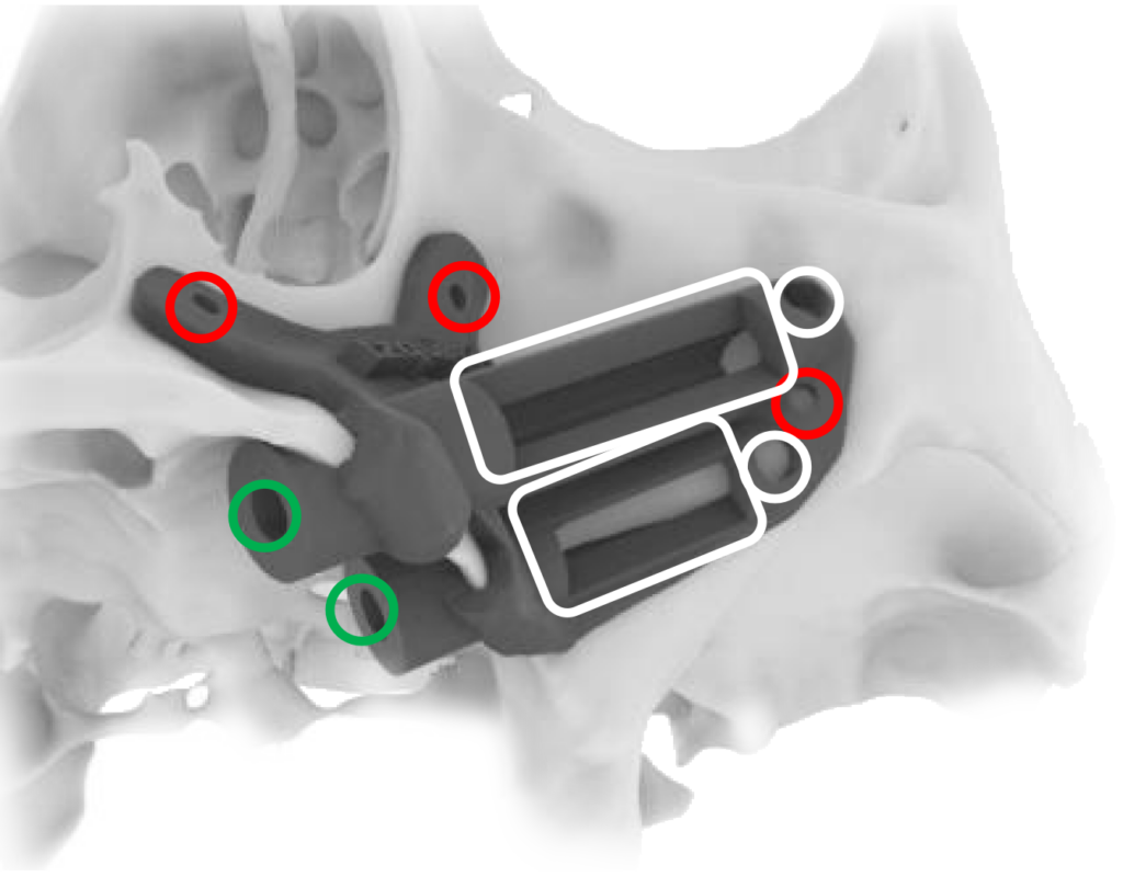
Characteristics of the guides
Red: Osteosynthesis fixation screws.
Green: Guided tubes.
White: Safety window introducing drill in the malar bone.
Rectangular windows: Verification of drilling.
Zygoma and pterygoid surgical guides
A CT scan allows planning and fabrication of surgical guides and the patient biomodel for precise, safe and personalized guidance of zygomatic and pterygoid treatments
When you access “UPLOAD TC” you must drag the folders or digital files you want to send to the indicated section of Smash.
When you're finished, we'll email you the details of the treatment record you just completed.
Within 24 to 48 hours, our engineers will confirm receipt of the digital files. If they are correct, they will proceed with pre-planning the processing according to your instructions.
They will then send you the prior planning by email, which you must approve or request a remote control meeting to verify it.